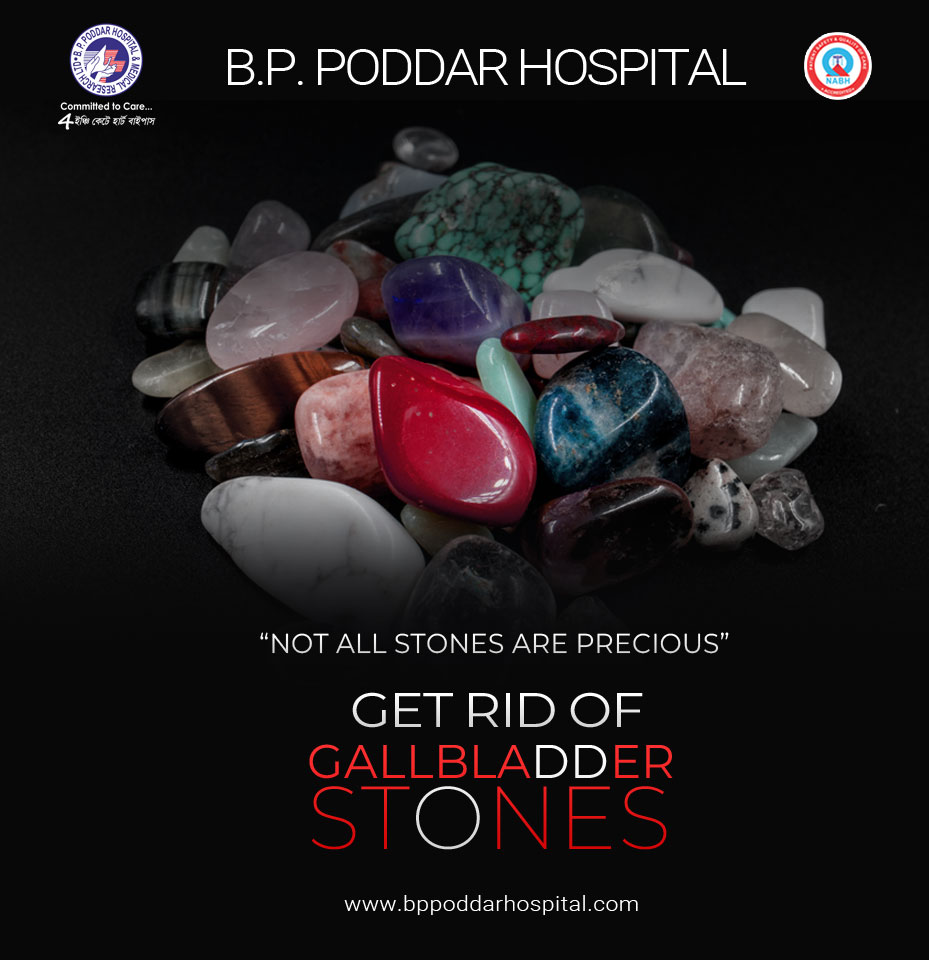
Gallbladder Stone- Know More
22 July, 2022
Gallstones, a very
prevalent issue has arisen in modern society as a result of lifestyle changes,
the intake of junk food, and irregular eating patterns. The development of
these stones usually starts in the gall bladder and sometimes these stones
start coming upwards towards the common bile duct along with the secretion of
bile during the digestion process. Usually, there is no onset of symptoms and these
issues are sometimes mistaken for indigestion, which is a transient condition
but if the size of the gallstones increases in number and size for an extended period,
then this leads to the severity and might worsen the condition of the patient
is not detected early.
What
are Gall bladder and Gallstones?
The gall bladder is a little,
pear-shaped structure situated beneath the liver on the upper right side of the
belly. It holds the bile that the liver produces for digestion and releases it
into the duodenum through the bile duct. Water, cholesterol, bile salts, and
pigments like bilirubin and others make up this bile. The breakdown of lipids
in our bodies is aided by bile.
Gallstones or Cholelithiasis which originate in the
gallbladder or bile ducts is solid masses resembling pebbles made of bile
precipitates. They range in size from the size of golf balls to small sand
grains. Gallstones are usually of two different types:
- Cholesterol stones- 80% of the
gallstones are of this type and these are usually yellowish-green in color.
- Pigment
stones- these gallstones are usually smaller and darker in
color. The color of these gallstones is determined by the excess production of
the bile pigment bilirubin produced by the liver.
What
are the causes? – There is no exact cause for the occurrence
of gallstones, still, some of the causes which lead to the formation of gallstones
are listed below:
- Due to a bad lifestyle such as extreme
junk food consumption rich in cholesterol regularly, skipping meals or eating
irregularly, severe consumption of alcohol and smoking, etc.
- Excess production of the bile pigment
bilirubin by the liver in cases of cirrhosis, other liver disorders, and blood
infections.
- Self-medication like regular intake of
NSAIDs to relieve chronic pain.
- Bile reflux- It is a reflowing action when
the bile bypasses the small intestine and instead rushes back into the stomach.
This condition usually leads to ascending the gallstones towards the common bile
duct from the gall bladder and finally results in obstructive jaundice due to
the blockage at the bile duct.
- People over the age of 40 years, obese or
overweight, and diabetic people are at higher risks for cholelithiasis.
What are the common symptoms observed?-
- Sudden intense abdominal pain usually at
the upper right portion usually right after the consumption of meals.
- Vomiting
- Indigestion, gas formation, and heartburn.
- Pain in the right shoulder, between the
shoulder blades, or towards the back.
- Jaundice-like symptoms like yellowing of
the skin and white portion of the eye.
- Dark-colored red urine and pale stool.
- Fever and chills
- Loss of appetite
- Severe complicated causes include
inflammation of the gall bladder (Cholecystitis), Pancreatitis, and
inflammation of the bile ducts (Cholangitis).
What
are the Diagnostic tests and Treatments available?-
- Pathological
examinations such as Blood tests and urine culture.
- Upper
abdomen ultrasonography- it is a non-invasive test to check the location of the
gallstones. However, only large-sized gallstones and the stones present in the gall
bladder are visible in this diagnostic test. Smaller-sized gallstones like
pigment stones and the gallstones present in the bile ducts are undetected, thus
it is not an accurate test.
- An
MRI technique called Magnetic Resonance cholangiopancreatography (MRCP)
especially visualizes the bile ducts. It produces highly detailed images of our
biliary system including the common bile duct and is non-invasive. MRCP
provides a clear picture and also rules out the possibility of tumors or
cancer. Depending upon the number, size, and location of the gallstones, the
doctor finally decides which surgical procedure needs to be performed on the patient
for the removal of the gallstones.
- Endoscopic
retrograde cholangiopancreatography (ERCP) is a more advanced and more non-invasive
procedure that is effective in locating gallstones if any of them are lodged in
the ducts and can also be removed using this technique. It is very useful in
the detection of smaller-sized pigment gallstones. It employs a combination of
X-rays and endoscopy which entails inserting a small camera into the upper gastrointestinal
(GI) system through the mouth.
- What is ERCP?
ERCP
is both diagnostic as well as a therapeutic technique for gallstones which
consists of an endoscope inserted through the mouth with an attached camera that
passes through the food pipe and reaches the top of the small intestine, then
the technician inserts another smaller tube into the first one to reach further
down into the bile ducts. They will inject a special dye through the tube and
then the video X-rays (fluoroscopy) are taken as the dye travels through the
ducts. Through ERCP, a clamp-like tool is inserted through the tube to remove
the stones which are present in the duct. The large-sized gallstones are
removed by lithotripsy in which the gallstones are first crushed or broken down
into smaller pieces and then each piece is taken out through the scope. The 190
series ERCP (Endoscopic Retrograde Cholangiopancreatography) is cutting-edge
technology in the earliest identification of biliary tract neoplasm,
cholangiocarcinoma, and pancreatic ductal adenocarcinoma.
- Endoscopic
Ultrasound- this combines both ultrasound and ERCP for the detection of
gallstones.
Cholecystectomy-
is laparoscopic keyhole surgery for the removal of the gall bladder when the
gallstones are accumulated in this sac. It is an ICG Enhanced Fluorescence
Guided procedure that requires a small incision, high resolution, minimal blood
loss, and a very low risk of infection.
- Medications
as prescribed by the medical practitioner.
Prevention
and Control-
- Avoid consumption of oily, fried, and
spicy foods.
- Intake of a healthy diet rich in high-
fiber such as fruits, vegetables, and whole grains.
- Consuming several little meals throughout
the day.
- Reduction of alcohol consumption and
caffeine.
- Management of stress.
- Maintaining a healthy weight, exercising,
etc.